Mohammed Obadi,
Ke-Xue Zhu,
Wei Peng,
Al-Farga Ammar,
Hui-Ming Zhou ![]()
For correspondence:- Hui-Ming Zhou Email: obadialariki@gmail.com Tel:+8618861825344
Received: 10 May 2016 Accepted: 14 September 2016 Published: 31 October 2016
Citation: Obadi M, Zhu K, Peng W, Ammar A, Zhou H. Effect of ozone gas processing on physical and chemical properties of wheat proteins. Trop J Pharm Res 2016; 15(10):2147-2154 doi: 10.4314/tjpr.v15i10.13
© 2016 The authors.
This is an Open Access article that uses a funding model which does not charge readers or their institutions for access and distributed under the terms of the Creative Commons Attribution License (http://creativecommons.org/licenses/by/4.0) and the Budapest Open Access Initiative (http://www.budapestopenaccessinitiative.org/read), which permit unrestricted use, distribution, and reproduction in any medium, provided the original work is properly credited..
Purpose: To investigate the effects of ozone treatment on chemical and physical properties of wheat (Triticum aestivum L.) gluten, glutenin and gliadin.
Methods: Wheat proteins isolated from wheat flour were treated with ozone gas. The physical and chemical properties of gluten proteins were investigated after treatment with ozone gas, with 5 g/h produced as a function of time (0, 30, and 60 min) in the study. To check whether the process of ozonation promoted changes in the quality of gluten proteins, sulfhydryl groups (SH), differential scanning calorimetry (DSC), secondary structure, SDS-PAGE, and rheology analyses were performed.
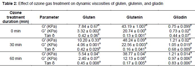
Results: Sulfhydryl group contents of wheat proteins ranged from 1.1 to 7.12 µmol/g. Sulfhydryl group content for all ozonated proteins was significantly lower than that of the control samples. Gluten proteins showed reduced SDS-PAGE band intensities of both high (HMW) gluten and glutenin subunits with increasing ozone gas treatment. The denaturation temperatures (Td) of ozonated gluten proteins were higher (99.80–106.79 °C) and the enthalpies of the ozonated gluten proteins were lower than those of the control samples. The storage moduli (G') and loss moduli (G”) of gluten and glutenin tended to increase from 7.84 to 10.20 KPa and 43.19 to 48.28 KPa, and from 3.33 to 4.06 KPa and 20.74 to 22.56 KPa, respectively, as ozone exposure increased from 0 to 30 min.
Conclusion: Ozone gas can oxidize wheat proteins. Exposing wheat proteins to ozone gas for an extended time (60 min) deteriorated wheat protein quality.
Introduction
Wheat (Triticum aestivum L.) gluten is very important for bread making. Grain proteins are classically divided into two fractions. Gliadins are present as monomers that either lack disulphide bonds or contain only intra-chain bonds; they provide cohesion and are responsible for the extensibility of gluten. Glutenins are polymeric, with high molecular weight, and they form complex polymers stabilized by inter-chain disulphide bonds. Glutenins contribute to the elasticity of gluten [1]. Ozone is one of nature's most powerful oxidants. It serves as a disinfectant in water treatments, destroys storage pests, and degrades mycotoxins to treat foods by sterilization [2]. Ozone has been used in the food processing industry, and is affirmed as generally recognized and safe (GRAS) [3]. The literature has demonstrated the efficacy of ozone for preservation of food grains. Tiwari et al [4] and Naito et al [5] demonstrated that ozone-treated grains at high concentrations (> 50 ppm) caused oxidative damage in cereal grain flours. Oxidation of sulfhydryl groups to disulfide bonds increases unextractable polymeric protein content [6]. There are growing areas of applications of ozone in the industry, but a limited amount of research has focused on using ozone as an oxidant of wheat proteins. The extent to which oxidation affects the physical and chemical properties of wheat proteins is unknown. In this study, the physiochemical properties of proteins (i.e., gluten, glutenin, and gliadin) isolated from non-ozone-treated flour were examined by studying chemical characteristics using SDS-PAGE, free SH groups, differential scanning calorimetry (DSC) parameters, secondary structure, and rheological measurements.
Methods
Preparation of gluten, glutenin, and gliadin
Wheat flower with medium protein content was obtained from the China Oil and Foodstuffs Corporation Co., Ltd. The protein content was 10.20 % on a dry basis. This flour was not bleached and had no added ingredients. A 300 g flour sample was mixed with NaCl solution (0.4 M, 160 mL) in a Farinograph. Then, after several minutes of resting time, the dough was washed manually with an NaCl solution (0.4 M, 3 L) until a cohesive mass (gluten) was obtained. The crude gluten was washed with distilled water to remove NaCl, followed by centrifugation at 5000 rpm for 10 min. Gluten (20 g) was shaken with dichloromethane (300 mL) for 1 h at room temperature to separate the glutenin and gliadin, filtered through filter paper, and filtered with Whatman filters. The procedures described above were repeated three times. Then, the gluten was dried at room temperature. Gliadin was extracted from 20 g of a 60 % gluten sample for 30 min, vortexed for 1 min every 10 min, and centrifuged for 10 min at 3000 rpm. Extraction was performed three times. The supernatants were pooled, and ethanol was removed using a rotary evaporator at 30 °C. Prior to the second and third extraction steps, the cohesive glutenin was mechanically disrupted with a spatula. The extraction was performed at 20 °C for 3 h, with centrifuging (4000 rpm, at 4 °C for 10 min) after each extraction. Following ethanol extraction, the gliadin and glutenin sediments were freeze-dried.
Ozone treatment
Proteins (gluten, glutenin, and gliadin) isolated from non-ozone-treated flour were treated with ozone gas using an ozone generator (model OJ-8003k-A, Guangzhou, China) with production of 5 g/h. After freeze-drying the gluten, the glutenin and gliadin samples were placed in a reaction vessel (rotating vessel designed for an evaporator) connected to the ozone generator. The samples were exposed to ozone gas for 0, 30, and 60 min. Air flow was set at 5 L/min. All samples were prepared in triplicate.
SDS–PAGE (sodium dodecyl sulfate-polyacrylamide gel electrophoresis) analysis
Sodium dodecyl sulfate-polyacrylamide gel electrophoresis (SDS-PAGE) analysis was performed on 10 % polyacrylamide gel. Proteins were separated in a 1-mm-thick preparative gel consisting of 4 % stacking gel and 12 % resolving gel. The gluten and glutenin samples (5 mg) were extracted with 1 mL of a non-reduced sample buffer solution containing 0.125 M Tris-HCl, 2 % (w/v) SDS, 10 % (v/v) glycerol, and 0.01 % w/v bromophenol blue at a pH of 6.8.
The protein–buffer mixtures were vortexed for 2 min and allowed to stand at room temperature for 2 h. Then, they were centrifuged at 35,714 rpm for 20 min at 4 °C, and the supernatant was heated for 3 min at 90 °C in a boiling water bath. After cooling to room temperature, the samples (50 µL) were loaded onto the gels. For reduced proteins, the sample buffer contained 5 % (v/v) β-mercaptoethanol. Electrophoresis was conducted at room temperature, with constant voltage at 100 V.
Determination of free SH groups
To determine the concentration of free sulfhydryl (SH) groups in gluten proteins, the following solvents were used: Tris-glycine-EDTA (TGE-EDTA; 10.4 g of Tris, 6.9 g of glycine, and 1.2 g of EDTA per liter, pH = 8.0, denoted as TGE buffer); 2.5 % SDS in TGE (SDS-TGE); and Ellman’s reagent (5,50-dithiobise2-nitrobenzoic acid, DTNB) in TGE (4 mg/mL). The 40 mg of samples were incubated with 4 mL of SDS-TGE for 30 min and vortexed every 10 min. Then, 0.04 mL of Ellman’s reagent was added and mixed in a 25 °C water bath for 30 min to prevent a light reaction.
Absorbance values were converted to concentrations of free SH, using a calibration curve with reduced glutathione. The solution was then subjected to centrifugation at 5000 rpm for 15 min, and the absorbance of the supernatant was measured at 412 nm against the blank sample, which lacked Ellman’s reagent and a sample.
Differential scanning calorimetry (DSC)
Differential scanning calorimetry (DSC) analysis was conducted using a TA Q100-DSC thermal analyzer (TA Instruments, Newcastle, DE, USA) following [7]. Approximately 2.0–3.0 mg of protein samples were weighed in aluminum pans, hermetically sealed, and heated in the DSC from 20 to 150 °C at 5 °C/min using N gas at a flow rate of 80 mL/min. An empty pan was used as a reference. The denaturation peak temperature (Td) and enthalpy (∆H) were obtained, and the half-peak height and width of endothermic peaks were obtained from thermograms using Universal Analysis 2000 (version 4.1D software; TA Instruments Waters, LLC, Newcastle, DE, USA).
Circular dichroism (CD) spectroscopy
A 0.1-mg/mL sample of a gliadin fraction solution in 50 % (v/v) ethanol was analyzed using a CD spectrophotometer (Biologic Science Instruments, Grenoble, France). The light path was adjusted (190–250 nm) using a 0.1-cm quartz cuvette [8].
Fourier transforms infrared (FTIR) spectroscopy
Fourier transforms infrared (FTIR) spectra were recorded on an FTIR spectrophotometer (NEXUS, Thermo., US) with single-reflection diamond attenuated total reflection (ATR) and mercury-cadmium-telluride (MCT) detectors. Spectra of an empty cell were used as a background, and cells were recorded for 64 scans at 4-cm-1 resolution. Hydrated samples were placed on the ATR crystal, and the spectra were recorded under the same conditions as the background. Two sub-samples were used for each determination. Experiments were conducted at room temperature using OMNIC software (version 6.1a, Thermo Nicolet Corp.) to evaluate secondary structural changes in the protein [9].
Rheological studies
Dynamic rheological measurements were recorded at 25 °C with a Rheometer AR 1000 (TA Instrument, Newcastle, DE) using parallel-plate geometry (40-mm diameter, 1-mm gap). Protein samples were sheared at 0.2 % strain through a frequency sweep from 0.1 to 10 Hz and at a constant temperature of 25 °C. Hydrated gluten and glutenin samples were pre-shaped to disks (25 mm diameter, 5 mm thickness), relaxed for 30 min, and fixed to the parallel plates of the stress rheometer coated with thin films of cyanoacrylate. The edges of the samples were covered with paraffin oil. The 3-mm gap was adjusted within 5 min, followed by a final relaxation of 10 min before measurements were recorded.
Statistical analysis
Each experiment was performed in triplicate, and the means and standard deviations (SDs) were calculated. SPSS 16.0 for Windows software was used for statistical analysis. Analysis of variance (ANOVA) was followed by Duncan’s multiple comparison test to assess differences. The significance level was defined as p < 0.05.
Results
Protein profile
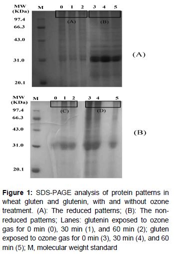
To better understand this phenomenon, both reduced (A) and non-reduced (B) SDS-PAGE were performed to characterize the changes in gluten and glutenin proteins treated with ozone gas. Ozone gas had no effect on these proteins. The protein-reducing patterns are shown in B. There were no major differences in most of the visible bands among all samples.
Ozone gas effect on SH groups
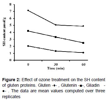
Ozone gas has been used to promote the oxidation of sulfhydryl groups, which results in the formation of disulfide bonds among glutenin molecules [10]. Gliadins are monomers with intra-chain disulfide bonds, whereas glutenin polymers have both intra and inter-chain disulfide bonds. Disulfide bonds are known to play an important role in maintaining the gluten network structure. Gluten depolymerized by disulfide bond breakage was used to increase the free SH content [11]. Free SH group exposure was monitored as a function of treatment time. The free SH group content in wheat gluten, glutenin, and gliadin decreased significantly as ozone treatment time increased (). This was likely due to the disulfide bonds formed in these proteins in response to ozone treatment. The SH content in gluten was reduced from 7.12 to 4.88 µmol/g with increased ozone gas treatment (). The SH content of glutenin was reduced from 4.20 to 2.50 µmol/g, and that of gliadin from 2.04 to 1.10 µmol/g with increasing ozone gas treatment.
Thermal properties
To investigate the effect of ozone gas on thermal properties of gluten proteins, samples were thermally scanned in a DSC. The denaturation temperature and enthalpy ∆H are the main parameters of the DSC profile. They occur during the baking process and ‘setting’ of the gluten network, significantly affecting the characteristics of the baked products [12]. In the present study, the results showed a slight increase in the denaturation transition temperature (Td) of gliadin compared to samples not treated with ozone ().
Secondary structure
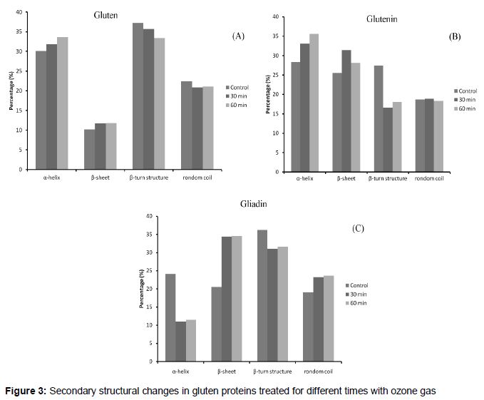
Far-UV circular dichroism (CD) spectroscopy was used to study the conformation of gliadin exposed to ozone gas [13]. FTIR spectroscopy is a useful tool to characterize protein secondary structures, with precision between the purely predictive and molecular coordinate approaches [14]. FTIR spectroscopy was used to determine secondary structures of gluten and glutenin viscoelastic bodies. The amide I band (1600–1700 cm-1) in FTIR spectra was mainly correlated with the secondary structure. We investigated how ozone treatment changed the structure of wheat gluten. As shown in , the gluten and glutenin secondary structures in the control sample were 30.1 % and 28.3 % of the α-helix, 10.2 % and 25.5 % of the β-sheet, and 22.4 % and 18.7 % of the random coil for gluten and glutenin, respectively. After ozone exposure for 60 min, 33.6 % and 35.6 % of the α-helix, 11.8 % and 28.09 % of the β-sheet, and 21.1 % and 18.3 % of the random coil were observed in gluten and glutenin, respectively.
Rheological properties
Gluten plays a major role in the rheological properties of flour dough that are important for the quality of bakery products. The averages of three measurements of elastic modulus G', viscous modulus G”, and Tan δ for the gluten, glutenin, and gliadin in samples that had been ozonated for different times are shown in . Gluten and glutenin had predominately viscoelastic solid behavior, whereas gliadins mainly conferred viscous flow, indicating dominance of the elastic characteristic [15]. Elastic modulus G' and viscous modulus G″ of gluten and glutenin in samples exposed to ozone for varying times are presented in . The gluten and glutenin of protein samples exposed to ozone for 30 min exhibited increased modulus G' and viscous modulus G″ compared to the control.
Discussion
These SDS-PAGE results are in agreement with the findings of Li et al [16], which showed that ozone treatment had no effect on the band intensities in reduced SDS-PAGE patterns. In contrast to the proteins, significant effects on gluten- and glutenin-ozonated samples were observed for the non-reduced SDS-PAGE patterns. The effect was particularly noticeable in terms of the intensity of bands within the range of 31 – 45 kDa in ozone-treated glutenin. In addition, an increase in the intensity of bands was observed in glutenin. A very remarkable reduction in band intensity was observed in gluten samples exposed to ozone for 60 min. These results indicate that protein treated with ozone for extended periods might cause scission of the disulfide bond (S–S bonds); this would help to break down the protein structure, and the labile peptide and amide bonds would be ruptured. Oxidation from ozone treatment of gluten and glutenin (60 min) caused partial depolymerization of high-molecular-weight proteins and increased content of low-molecular-weight protein polymers.
The increase in disulfide bonds improves the gluten matrix by sulphydryl group oxidization, thus increasing the cross-linking of protein polymers. Glutenin subunits were polymerized with inorganic oxidization using K iodation, K bromation, and hydrogen peroxide, as described by Veraverbeke et al [17,18], and molecular oxygen [19]. This polymerization was due to the oxidation of SH groups. Cataldo [20] reported that ozone oxidizes cysteine in protein to form cystine disulfide bonds. The results from our study were similar to those reported by Chittrakorn [10], who also found that SH groups were oxidized during chlorine treatment. Ozone treatment oxidized sulfhydryl groups to disulfide bonds, which could explain the increased dough strength [6].
These values are similar to those reported by Sinha et al [21], who stated that chlorination caused a slight increase in denaturation of gliadin. Myers [22] reported that large increases in denaturation (Td) values are expected for proteins with a high proportion of hydrophobic residues involved in the denaturation mechanism. In the present study, the Td values of ozone-treated and non-ozone-treated samples were not significantly different. However, ozone-treated samples had slightly higher Td values, indicating an increase in the hydrophobicity of ozonated samples. Falcao-Rodrigues et al [16] proposed that the denaturation of gluten proteins is accompanied by decreased solubility, and proceeds to a point where the gas vesicle walls are fixed and expansion ends. Thus, the higher Td of gliadins treated with ozone would result in large gas cells and higher loaf volume in bread. The results from the DSC analysis showed that ozone treatment decreased the enthalpies ∆H of gluten proteins. The reduction in enthalpies ∆H of ozone-treated proteins might be due to ozone gas, which caused unfolding of gluten proteins, disrupting the ordered structure during treatment. Sinha et al [21] also found a decrease in the enthalpy ∆H value in of chlorinated gliadin samples. Deterioration of gluten proteins caused disruption in the ordered structure, which could weaken baked goods.
These results suggest that β-sheet and α-helix are sensitive to the action of ozone. Changes in the content of β-sheet and random coil structures of proteins exposed to ozone treatment affect intermolecular hydrogen bonding of the β-sheets. Misra NN et al and Safonova et al [23,24] reported that ozone gas affected the secondary structure of wheat flour proteins based on FTIR spectroscopy results. The CD spectra calculation indicated a significant decrease (approximately 12 %) in the α-helix for gliadin samples in response to increased ozone gas treatments. Both the β-sheet and random coil increased by nearly 14 and 4 %, respectively with increased ozone gas treatments. Enhancement of the α-helix structure had detrimental effects on the β-sheet and random coil structures. The effects on the secondary structure caused some conformational changes in ozone-treated wheat gluten. New intermolecular cross-links would appear to cause joining within two molecular proteins and changes in the secondary structures.
After 30 min of exposure to ozone, gluten and glutenin of the protein samples displayed decreased elastic modulus G' compared with control samples. This decrease might be attributed to the reduced functional properties of protein due to oxidation by ozone exposure. This was confirmed by our SDS-PAGE analysis of gluten proteins, where overexposure of gluten proteins to ozone caused partial depolymerization of high-molecular-weight proteins and increased the content of low-molecular-weight protein polymers.
Ozone may have influenced more protein molecules with increased exposure to gas ozonation, causing an extensive change in the protein structure, which appeared as differences in rheological properties. Tan δ is generally an indicator of protein quality [25]. Tan δ gives a measure of the viscous and elastic response of the material being tested. Tan δ values of ozonated gluten and glutenin increased compared to the control. However, further treatment of gluten and glutenin with ozone gas (60 min) increased tan δ (). These changes might be due to the deteriorating protein quality. This was probably attributable to the depolymerization of GMP in gluten and glutenin due to overexposure of proteins to ozone. Over oxidation of proteins would make the dough too weak and susceptible to expanding during baking. It is also hypothesized that excess formation of disulfide bonds over longer periods of time, with the accompanying loss of sulfhydryl groups, might hinder disulfide bond interchange, leading to more rapid breakdown of dough. An earlier study suggested that glutenin polymerization leads to a significant increase in the elastic modulus of the network [26]. A significant correlation exists between viscoelastic properties in the dough and the amount of the glutenin macro polymer (GMP) [27]. Ozone could be used to improve protein quality and change rheological properties to make the dough more elastic.
Conclusion
Ozone gas treatment has been applied to gluten, glutenin, and gliadin samples to estimate the nature and extent of changes in gluten protein properties. Overall, the findings revealed that exposing gluten proteins to ozone gas for a short period of time (30 min) enhanced its physical and chemical properties (SDS-PAGE, SH, DSC, secondary structure, and rheology properties) than longer term (60 min) exposure to ozone gas.
Declarations
Acknowledgement
References
Archives


News Updates